- Home
- Live-Cell Analysis
- IncuCyte® CytoLight Reagents
IncuCyte® CytoLight Reagents
IncuCyte® CytoLight Reagents enable efficient, non-perturbing, cytoplasmic labeling of living cells. They are a unique range of lentiviral-based labeling reagents with simple transduction protocols that enable expression of a cytoplasmically soluble green (GFP) or red (mKate2) fluorescent protein in your choice of primary, immortalized, dividing, or non-dividing cells.
These labeling reagents have been validated for use with the IncuCyte® Live-Cell Analysis System and are ideal for monitoring and quantifying the real-time formation of multicellular structures and cell-cell interactions in co-culture systems.
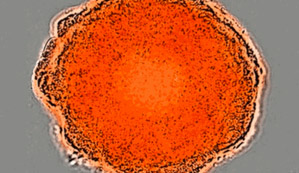
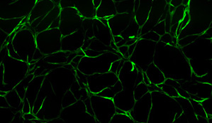
Label cells without altering cell function and monitor live-cell interactions over days and weeks. 3D-spheroid formation and growth using A549 tumor cells labeled with IncuCyte® CytoLight Red Reagent (left) and vascular tube formation in a co-culture model of angiogenesis using HUVEC primary endothelial cells labeled with IncuCyte® CytoLight Green Reagent (right). Note that cells remain healthy and fluorescence expression is maintained over the entire time course (>10 days).
IncuCyte® CytoLight Reagents – IncuCyte® Publications
Researchers around the world are using IncuCyte® CytoLight Reagents to monitor and quantify the formation of multicellular structures in complex culture models. Explore the IncuCyte® publications library and learn why IncuCyte® technology has been sited in over 700 peer reviewed publications.
Phosphoproteomic analysis implicates the mTORC2-FoxO1 axis in VEGF signaling and feedback activation of receptor tyrosine kinases.
Zhuang G, Yu K, Jiang Z, Chung A, Yao J, Ha C, Toy K, Soriano R, Haley B, Blackwood E, Sampath D, Bais C, Lill JR, Ferrara N.
Science Signaling 6 (271):ra25 (2013);
Researchers at Genentech used HUVEC cells transduced with the IncuCyte® CytoLight™ Green reagent in a co-culture model of angiogenesis to reveal a link between FOXO1 inhibition of VEGF signaling and the PI3K-mTORC2 signally axis.
Label and monitor cell-cell interactions without perturbing function or morphology
- Monitor the assembly of multicellular structures in real time over days or weeks
- Non-perturbing to cell biology and function
- Simple labeling protocols – compatible with your choice of cells
- Validated for use with the IncuCyte® Live-Cell Analysis System
- Ideal for co-cultures and multiplexing with IncuCyte® apoptosis or cytotoxicity assays

IncuCyte® CytoLight Lentivirus Reagents
- Long term, homogeneous expression
- Simple ‘seed-transduce-select’ protocols
- Ideal for generating stable cell populations
IncuCyte® CytoLight Lentivirus Reagents
| Name | Type | Promoter | Selection | Ex. maxima | Em. maxima | Catalog No. |
|---|---|---|---|---|---|---|
| IncuCyte® CytoLight Green | Lentivirus | EF-1α | Puromycin | 483 nM | 506 nM | 4481 |
| IncuCyte® CytoLight Red | Lentivirus | EF-1α | Puromycin | 588 nM | 633 nM | 4482 |
| IncuCyte® CytoLight Green | Lentivirus | CMV | None | 483 nM | 506 nM | 4513 |
IncuCyte® CytoLight Pre-labeled Cell Lines
Direct access to cells stably expressing NucLight™ Green or Red fluorescent proteins. No need to transduce. Simply thaw and use.
| Name | Selection | Volume | Ex. maxima | Em. maxima | Catalog No. |
|---|---|---|---|---|---|
| HUVEC CytoLight Green Cells | None | 1.7x10⁵ cells/vial | 483 nM | 506 nM | 4453 |


