- Home
- Live-Cell Analysis
- IncuCyte® NucLight Reagents
Incucyte® Nuclight Reagents
Incucyte® Nuclight Reagents enable efficient, non-perturbing, nuclear labeling of living cells, using a range of different cell-labelling strategies. Our Lentiviral-based labeling reagents enable expression of a nuclear-restricted green (GFP), red (mKate2), orange (TagRFP) and NIR (iRFP713) fluorescent protein in your choice of primary, immortalized, dividing, or non-dividing cells. Alternatively, cells can also labeled with our Nuclight Rapid Red or NIR nuclear binding fluorescent dyes.
These versatile labeling reagents have been validated for use with the Incucyte® Live-Cell Analysis System and enable automated cell counting within your tissue culture incubator. The Incucyte® Nuclight live-cell labeling reagents are ideal for use in cell counting and proliferation studies and can be easily multiplexed with other Incucyte® Cell Health reagents for simultaneous measurements of apoptosis and cytotoxicity in monoculture or co-culture models.
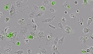
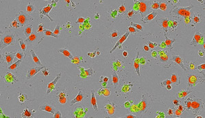
Label cell nuclei and accurately count living cells in real time without altering cell function. Time-lapse movie of co-cultured nuclear-labeled HT1080 cells (green nuclei, Nuclight Green) and A549 cells (red nuclei, Nuclight Red). Note the healthy cell morphology over the 72 hour period and the different doubling times of the two cell types which can be derived from the time vs. cell count plot (doubling times; HT-1080, 16 hours; A549, 20 hours)
Incucyte® Nuclight Reagents – IncuCyte® Publications
Researchers around the world are using Incucyte Nuclight Reagents to count living cells in relevent co-cultures and screening models. Explore the Incucyte® publications library and learn why Incucyte systems have been sited in over 2000 peer reviewed publications.
A co-culture genome-wide RNAi screen with mammary epithelial cells reveals transmembrane signals required for growth and differentiation.
Burleigh, A. et al. Breast Cancer Res. 17, (2015).
Researchers at the British Columbia Cancer Agency, Vancouver, Canada used Nuclight Red labeled mammary epithelial cells to perform a genome-wide siRNA screen in co-cultures. IncuCyte® proliferation (nuclei counts) and apoptosis assays were used to functionally identify genes regulating mammary epithelial cell growth.
Label and count living cells without perturbing cell function or morphology
- Count living cells in real time over days – calculate true doubling times and growth rates
- Non-perturbing to cell biology and function
- Simple labeling protocols – compatible with your choice of cells
- Validated for use with the Incucyte® Live-Cell Cell Analysis System
- Ideal for co-cultures and multiplexing with IncuCyte® apoptosis or cytotoxicity assays
Which Incucyte® NucLight Reagent should I choose?
- Incucyte Nuclight Lentivirus Reagents are ideal for generating homogeneous, stably expressing cell lines with long term, consistent fluorescence expression. Transduce your cells once and use for multiple studies or screening campaigns.
- IncuCyte Nuclight Rapid Reagents are ideal in monoculture scenarios where it may be difficult or undesirable to transfect cells with fluorescent proteins. These mix-and-read reagents are perfect when multiple cells lines require rapid and efficient labeling without the need to select for stable clones.
Use the table below to identify which NucLight Reagent suits your needs:
| Nuclight Lentivirus Reagents | Nuclight Rapid Reagents | |
|---|---|---|
| Rapid maximal expression < 20h | ||
| Expression duration > 72h | ||
| Use to make stable cell lines | ||
| Ideal for transient expression | ||
| Use under BSL1 conditions | ||
| Simple protocols |
* Duration of expression is cell type dependent. Expression will last longer than 72 hours in slowly dividing, or non-dividing, cells (e.g. neurons) and may last less than 72 hours in some rapidly dividing cell types.
Ordering Information
Incucyte® Nuclight Lentivirus Reagents
| Reagent | Type | Promoter | Selection | Volume | Ex. maxima | Ex. minima | SDS (US) | Catalog No. |
|---|---|---|---|---|---|---|---|---|
| IncuCyte® NucLight Green | Lentivirus | EF-1α | Puromycin | 0.2 mL | 483 nM | 506 nM | 4624 | |
| IncuCyte® NucLight Green | Lentivirus | EF-1α | Puromycin | 0.6 mL | 483 nM | 506 nM | 4475 | |
| IncuCyte® NucLight Red | Lentivirus | EF-1α | Puromycin | 0.2 mL | 588 nM | 633 nM | 4625 | |
| IncuCyte® NucLight Red | Lentivirus | EF-1α | Puromycin | 0.6 mL | 588 nM | 633 nM | 4476 | |
| IncuCyte® NucLight Green | Lentivirus | EF-1α | Bleomycin | 0.2 mL | 483 nM | 506 nM | 4626 | |
| IncuCyte® NucLight Green | Lentivirus | EF-1α | Bleomycin | 0.6 mL | 483 nM | 506 nM | 4477 | |
| IncuCyte® NucLight Red | Lentivirus | EF-1α | Bleomycin | 0.2 mL | 588 nM | 633 nM | 4627 | |
| IncuCyte® NucLight Red | Lentivirus | EF-1α | Bleomycin | 0.6 mL | 588 nM | 633 nM | 4478 |
IncuCyte® Nuclight Rapid Reagents
| Reagent | Type | Promoter | Selection | Volume | Excitation | Emission | Catalog No. |
|---|---|---|---|---|---|---|---|
| IncuCyte® NucLight Rapid Red | Rapid | N/A | N/A | 50 µL | 655 nm | 681 nm | 4717 |
IncuCyte® NucLight Pre-labeled Cell Lines
Our pre-labeled IncuCyte® NucLight cell lines enable direct access to cells stably expressing NucLight Green or Red fluorescent proteins. No need to transduce. Simply thaw and use.
| Reagent | Quantity | Selection | Ex. maxima | Ex. minima | Catalog No. |
|---|---|---|---|---|---|
| A549 NucLight Green Cells | 1x10⁶ cells/vial | Puromycin | 483 nM | 506 nM | 4492 |
| A549 NucLight Red Cells | 1x10⁶ cells/vial | Puromycin | 588 nM | 633 nM | 4491 |
| HeLa NucLight Green Cells | 1x10⁶ cells/vial | Puromycin | 483 nM | 506 nM | 4490 |
| HeLa NucLight Red Cells | 1x10⁶ cells/vial | Puromycin | 588 nM | 633 nM | 4489 |
| HT-1080 NucLight Green Cells | 1x10⁶ cells/vial | Puromycin | 483 nM | 506 nM | 4486 |
| HT-1080 NucLight Red Cells | 1x10⁶ cells/vial | Puromycin | 588 nM | 633 nM | 4485 |
| MCF7 NucLight Green Cells | 1x10⁶ cells/vial | Puromycin | 483 nM | 506 nM | 4528 |
| MCF7 NucLight Red Cells | 1x10⁶ cells/vial | Puromycin | 588 nM | 633 nM | 4524 |
| Neuro-2a NucLight Green Cells | 2x10⁶ cells/vial | Puromycin | 483 nM | 506 nM | 4511 |
| Neuro-2a NucLight Red Cells | 2x10⁶ cells/vial | Puromycin | 588 nM | 633 nM | 4512 |
| THP-1 NucLight Red Cells | 1x10⁶ cells/vial | Puromycin | 588 nM | 633 nM | 4612 |
| Jurkat NucLight Red Cells | 1.5x10⁶ cells/vial | Puromycin | 588 nM | 633 nM | 4613 |
Nuclight Protocols
Incucyte® Cell Transduction Protocol
This protocol provides an overview of the Incucyte® Cell Transduction Assay methodology. It is compatible with the IncuCyte® live-cell analysis system and enables real-time cell counting using your choice of cells and treatments. The Incucyte® Nuclight range of live cell labeling reagents are used to fluorescently label the nuclei of living cells without perturbing cell function or biology.
Download the Incucyte® Cell Transduction Protocol
IncuCyte® Nuclight Rapid Red Cell Labeling Protocol
This protocol provides an overview of the Incucyte Nuclight Rapid Cell Labeling Reagent methodology. It is compatible
with the IncuCyte live-cell analysis system and describes how IncuCyte Nuclight Rapid Red can be used to fluorescently
label the nucleus of living cells without perturbing cell function or biology. In addition, this reagent can be multiplexed with IncuCyte® Annexin V Green, Caspase-3/7 Green, or Cytotox Green reagents for simultaneous readouts of apoptosis or cytotoxicity in the same well.
Download the Incucyte® Nuclight Rapid Red Cell Labeling Protocol
Related Applications

Proliferation Assays
Measure label-free cell growth or growth inhibition and count living cells in real time

Apoptosis Assays
Detect apoptosis in living cells and in real time using simple mix-and-read protocols


